Z.U.M.J.Vol.20; N.1; Jan; 2014
Samer Badee a , Naslshah Kazem a , Hesham Khalid b
a Department of otorhinolaryngology, Faculty of medicine ,
Benha University, Egypt
b Department of Cardiology, Faculty of medicine , Benha University, Egypt
ABSTRACT
Introduction: Obstructive sleep apnea syndrome (OSAS) is characterized by repetitive partial and total collapse of the upper airway that induces stressful arousals throughout sleep to reestablish breathing. Hypertension was more frequently seen in patients with OSAS than in patients without OSAS and hypertension frequency increased in parallel to the severity of OSAS .The goal of uvulopalatopharyngoplasty (UPPP) in the treatment of obstructive sleep apnea syndrome is to reduce obstruction by eliminating redundant tissue in three areas: soft palate, tonsils and pharynx . Objective: The aim of this prospective study was to determine the relationship between the severity of OSA and primary hypertension by using acoustic pharyngometry pre and post uvulopalatopharyngoplasty.Study design: A prospective comparative study.Subjects and methods: 50 patients with both OSA and primary hypertension examined and investigated with acoustic pharyngometer, then operated up on with UPPP and reassessed for their blood pressure and acoustic pharyngometer after 3 months from the operation in comparson with 10 normal individuals.Results: The results 3 months after UPPP showed that the total number of cured and improved cases by acoustic pharyngometery were 9 & 23 cases (18% & 46%) & cases of pimary hypertension cured & improved were 8 & 40 cases that represented 16% & 40% respectively Conclusion:The severity of OSA can be determined objectively by using acoustic pharyngometry and blood
pressure in the sleep apnea patients might gradually decreased after UPPP surgery .
Keywords obstructive sleep apnea, primary hypertension,acoustic phyryngometry.
INTRODUCTION
Obstructive sleep apnea syndrome (OSAS) is characterized by repetitive partial and total collapse of the upper airway that induces stressful arousals throughout sleep to reestablish breathing. The prevalence has been reported as high as 20 % in the adult population 1 .
The pathophysiological basis of OSA is periodic airway occlusion resulting in sleep and blood gas disturbance which leads to cardiovascular disease, hypertension, arrhythmia, disturbed psychosocial performance, metabolic and neuroendocrine dysfunction and increased risk of traffic accidents2 . Acoustic pharyngometry is a modern diagnostic method based on physical principle of acoustic reflection. It is useful for volume analysis of oro- pharyngo-laryngeal spaces. It’s a clinically useful tool for localizing the narrowed portion of the upper airway and predicting OSA3
Hypertension was more frequently seen in patients with OSAS than in patients without OSAS and hypertension frequency increased in parallel to the severity of OSAS4 .
Grotz W et al, 2006 5 approved a causal relationship between sleep apnea and hypertension. Undiagnosed sleep apnea is probably the most important reason for essential hypertension and they advised that all hypertensive patients should be asked for snoring, breathing arrest and daytime sleepiness.
The goal of uvulopalatopharyngoplasty (UPPP) in the treatment of obstructive sleep apnea syndrome is to reduce obstruction by eliminating redundant tissue in three areas: soft palate, tonsils and pharynx 6 .
The aim of this study was to define the relationship between the severity of obstructive sleep apnea and primary hypertension by using acoustic pharyngometry and to determine if essential hypertensive patients will improve after treatment of OSAS by uvulopalatopharyngoplasty.
SUBJECTS AND METHODS
This prospective study conducted in Benha university hospital and Benha teaching hospital in the period between 1/3/2012 – 1/12/2012. We selected 50 patients with both primary hypertension without other co-morbidity factors, middle aged (30-50 years) of both sexes, non obese, non smokers or alcoholics at the ENT clinic, after taking a full detailed history of symptoms of OSAS(with participation by the bed partner if possible ) and examination by ENT surgeon, representing group (A).
We also selected 10 young healthy adults of both sexes as a control group, representing group (B). All controls gave no history of snoring, and hypertension was also verified negative for all individuals.
Members of group A were classified into 3 subgroups mild, moderate and severe grades sleep apnea according to American sleep association 2005 7 to
Mild grade included 17 patients. Moderate grade included 20 patients. Severe grade included 13 patients .
1. Body mass index calculated as weight in kg/ (height in meters2). Because obesity is defined as body mass index > 30 kg/m2, only individuals of BMI equal or less than 27 were included.
2. Collar size of less than 16.5 inches.
3. No apparent facial skeletal anomalies and normal dental occlusion.
4. ENT examination for signs suggestive of possible upper airway obstruction, such as deviated septum, hypertrophied turbinate or presence of polyps, enlarged tonsils, redundant soft palate, and webbing of the tonsillar pillars, abnormally huge tongue and the relationship of palatal arch to the tongue.
5. Blood pressure need to be recorded. Hypertension was defined as a systolic blood pressure (SBP) ≥ 140 mm Hg and/ or diastolic blood pressure (DBP) ≥ 90 mm Hg. Blood pressure was measured from the right arm of the seated participant after 5 minutes rest and was recorded to the nearest 2 mmHg using first and fifth Korotkoff sounds .Three blood pressure measurements were taken and the mean of the last two measurements was used in the analysis .All patients had neither received treatment for hypertension nor had withdrawn from treatment 3 weeks earlier. Patients were evaluated at the Internal Medicine Department to exclude the presence of secondary hypertension.
6-Those patients undergone acoustic pharyngometry to localize the site of obstruction and to determine the severity of OSA.
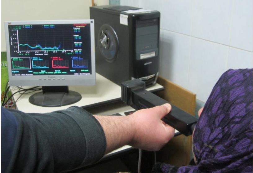
Procedure of acoustic pharyngometry: The device used was acoustic pharyngometer [E. Benson Hood Laboratories, Pembroke, Mass.USA]

Fig(1): Acoustic pharyngometry examination (Benha Teaching Hospital, ENT department, 2012).
A) Patient position: The subject is seated in a firm chair, straight back, adjustable seat height to maintain the head in neutral position and the wave tube in proper position.
B) Patient considerations: The test is done during normal quite breathing; therefore, the subject is allowed to sit down for a while during that time he is briefed about the test and its nature. Instruct the patient to stay still during the test, fix the gaze at a point on the opposite wall at the same gaze level. Patients were also told to think silently of “OOOh” in order to put the tongue in a relaxed position on the floor of the mouth and keep the velum closed as vowel phonation is through the mouth only.
C) Mouthpiece: The mouthpiece is made of rubber. It is designed to be placed with the teeth against the flange, biting down on the protruding tabs, and the lips over the flange to form acoustic seal.
D) Positioning of the wave tube: It is placed horizontally.
E) Operator: Some training on the operator himself or a volunteer prior to working with the equipment is helpful in order to get familiar with the equipment and consistently get a reproducible result. It is always important to watch the test setup rather than watching the computer screen.
Patients in group (A) were undergone UPPP which consists of the following steps:
Step 1:
Following orotracheal intubation and deep muscle relaxation, the mouth gag is positioned .This routinely provides adequate visualization and exposure. The amount of palate to be resected was determined by gently pushing the palate to the posterior pharyngeal wall and marking on its ventral surface the point where the palate met the posterior pharyngeal wall.
Step 2:
The tonsils, if present, are excised, and
excessive tissue along the posterior tonsillar pillar is removed. Posterior tonsillar pillars are sutured anteriorly to the anterior tonsillar pillar, which removes any excessive tissue and enlarges the oropharyngeal opening in the horizontal plane.
Determination of the correlation between ……….
The uvula also is excised, and a portion of the inferior edge of the soft palate frequently is removed. This area then is closed on itself, and the anterior wall of the nasopharynx, which is a posterior wall of the soft palate, is rotated anteriorly to enlarge the nasopharyngeal outlet into the pharynx. The resection of the uvula and portions of the palate enlarge the oropharynx in a vertical and a horizontal fashion, which results in an increase in the size of the oropharyngeal inlet. Three months after surgical treatment of patients with OSAS by uvulopalatopharyngoplasty, they undergone further measurement of the same parameters.
Statistical analysis was performed to show a significant difference between different parameters pre & post-operative.
RESULTS
Table (1): Statistical comparison between different grades of cases with OSA as regards the
extension and amplitude of OP segment:
Grade of OSA
Mild
Moderate
Severe
OP- extension Mean (cm)
3.7-3.9 3.8
4-4.2 4.1
4.3-4.5 4.4
OP-amplitude Mean (cm2)
0.9-1.4 1.15 0.5-0.8 0.65 0.2-0.4 0.3
Table (1): Shows the mean of OP segment amplitude and extension in different grades of cases with OSA. From this table we find that:
1. PatientswithmildOSA:theOPsegmentextensionisfrom3.7to3.9cm&theamplitudeisfrom0.9to1.4cm2.
2. Patients with moderate OSA: the OP segment extension is from 4 to 4.2 cm & the amplitude is from 0.5 to 0.8 cm2. 3. PatientswithsevereOSA:theOPsegmentextensionisfrom4.3to4.5cm&theamplitudeisfrom0.2to0.4cm2.
Fig (2) shows a pharyngogram of a normal person (control)
- Diagram: show normal (B, C, D, and E) waves.
- Amplitude of OP segment > 1.6 cm2.
- OP segment interval < 3.6 cm.
- Normal curve.
-114-
Z.U.M.J.Vol.20; N.1; Jan; 2014
Determination of the correlation between ……….
Fig (3): Shows a pharyngogram of a patient with redundant soft palate and hypertrophied elongated uvula (Severe OSA).
- Diagram: show normal (B) wave, marked depressed , elongated O-P segment (C) wave anddepressed (E) wave hypopharyngeal wave.
- Severe OSA as the amplitude of OP segment 0.2 cm2 and OP segment interval 4.5 cm.Table (2): Statistical comparison between group A (cases) and group B (controls) as regards acoustic pharyngometric data:
Variable Groups Mean
± SD
0.32 0.33
0.51 0.41
Student t test
15.27 10.68
P value 0.001 HS 0.001 HS
OP-amplitude (oropharynx)
OP- extension (oropharynx)
Table (2):
Case 0.69
Control 1.71
Case 3.95
Control 2.96
Shows the mean & the standard deviation (SD) of pharyngometric data of (cases & controls). It
shows also a highly significant difference in the oropharyngeal wave (C wave) amplitude and its extension
O-P segment (P<0.001).
Table (3): Statistical comparison between acoustic pharyngometric data for cases (group A) pre& post-operative :
Variable (case)
OP-amplitude (oropharynx)
OP- extension (oropharynx)
Table (3):
Groups Mean
pre op post op pre op post op
± SD Paired t test
P value
0.69 1.24 3.95 3.27
0.32 0.24
0.513 0.48
22.75 18.77
0.001 HS 0.001 HS
Shows the mean & the standard deviation (SD) of pharyngometric data of cases (pre& post-
operative). It shows also a highly significant difference in oropharyngeal wave amplitude and its extension
O-P segment (C wave) (P<0.001).
-115-
Z.U.M.J.Vol.20; N.1; Jan; 2014
Table (4): Outcome of cases 3 months after UPPP:
Determination of the correlation between ……….
outcome
Total
Acoustic pharyngometery
Total
Sleep index subjectively Blood pressure
cured 9 10 8
improved 23
not improved 18
Total 50
            21                  20
            19                  22
            50                  50
the oropharynx about (99%) of cases . Also this segment showed a highly significant difference between cases and controls. These results matched with Gelardi et al., (2007) 3 whostudied pharyngeal cross-sectional area by acoustic reflectometry found that an increased O-P segment extension was seen in patients with OSAS (98%). It is thus an indirect sign of excessive contact between soft tissues in the mouth and the pharynx. This common finding in OSAS patients underlines the etiological and pathological importance of existing stenosis in this anatomical region.
The American sleep association7 grades sleep apnea as follows: mild = 5-20 apneas per slept hour, moderate = 20-40 apneas per slept hour, severe > 40 apneas per slept hour. This method of grading is mainly subjective depending mainly on participation by the bed partner. Through our study, we found that subjects with OSA show differences in OP segment amplitude and extension according to the severity of the condition. Patients with mild OSA: the OP segment extension is from 3.7 to 3.9 cm & the amplitude is from 0.9 to 1.4 cm2. Patients with moderate OSA: the OP segment extension is from 4 to 4.2 cm & the amplitude is from 0.5 to 0.8 cm2. Patients with severe OSA: the OP segment extension is from 4.3 to 4.5 cm & the amplitude is from 0.2 to 0.4 cm2 (table 3). This study suggests that acoustic pharyngometry provides a useful parameter for determination of the severity of obstructive sleep apnea.
Ugeskr Laeger, 200910 established causal link between OSA and cardiovascular disease, in particular hypertension. The strong association between OSAHS and systemic hypertension has been well recognized11. The prevalence of hypertension in patients with OSAHS was reported as 45%12 and, even as high as 57.1% in middle-aged OSAHS patients13,14 demonstrates that nocturnal hypoxemia and augmented sympathetic nerve tension following repetitive
Table (4): Shows that the total number of cured cases was 9 with a percentage of 18%.While the number of cases that improved was 23 with a percentage of 46%.On the other hand, the total number of cases that not improved was 18 with a percentage of 36%.
* The patient was considered cured when his BP returned to normal level (SBP < 140 mmHg , DBP <90 mmHg), AI <5 per slept hour and normal acoustic pharyngometric data (normal OP segment amplitude and extension).
* The patient was considered improved when his BP, AI and acoustic pharyngometric data were better than pre-treatment but didn’t return to normal levels.
* The patient was considered not improved when his BP, AI and acoustic pharyngometric data were not affected by treatment.
DISCUSSION
OSA is a complex disease process characterized by repeated upper airway (UA) obstruction during sleep. The etiology is multifactorial and not fully understood.
Acoustic reflection technique is not a newly developed method. It was originally described by Jackson et al.,(1977)8, and further modified by Fredberg et al.,(1980), 9 who used this technique to measure tracheal cross sectional areas in human subjects. The technique is based on sending acoustic impulses along the respiratory tract. As they travel through the airways, they are partially reflected whenever there is a change in the airway cross-sectional area. By calculating the amplitude and temporal changes in the reflected pulse compared with the incident pulse, it is possible to assess cross-sectional area and volume from the oral cavity to hypopharynx. Through many studies the accuracy of pharyngometry-derived measurements has been evaluated.
From this study, it was found that the O-P segment (C wave) is the site most affected in patients of OSA. By far the most common stenosed site in cases of OSAS in our locality was
-116-
Z.U.M.J.Vol.20; N.1; Jan; 2014
hypoxia plays an important role in the etiology of systemic hypertension in patients with OSAHS.
Uvulopalatopharyngoplasty is the most commonly performed surgical procedure as treatment for OSAHS, which was first reported by Fujita et al, 198115.The goal of UPPP in the treatment of OSAHS is to reduce obstruction by eliminating redundant tissue in three areas: soft palate, tonsils and pharynx 6. In this study, we selected the patients that had an obstruction in their oropharynx and we attempted to remove the site of upper airway obstruction with UPPP surgery.
Yu S et al, 201016 studied the value of the surgery of revised uvulopalatopharyngoplasty on ambulatory blood pressure (BP) in (OSAHS) patients with hypertension and oropharyngeal obstruction. A retrospective cohort study was performed in 29 patients with OSAHS and hypertension, who received treatment with revised UPPP surgery. After surgery 1 month, the Apnea- hypopnea index (AHI) significantly decreased (P < 0.05). In the meantime, compared to pretreatment, 24-h DBP, and 24-h SBP decreased significantly (P < 0.05).In all 29 patients, 6 patients’ BP decreased to normal (20.6%); other patients’ BP decreased from pre-treatment, but didn’t decrease to normal. In our study, 9 patients’ BP decreased to normal (18%); 23 patients’ BP decreased from pre-treatment, but didn’t decrease to normal (46%); while there were 18 patients’ BP didn’t decrease after surgical treatment (36%). In our study; after surgery 3 months, the Apnea- hypopnea index (AHI) Shows a highly significant difference (P<0.001). In the meantime, compared to pretreatment, 24-h DBP, and 24-h SBP Shows a highly significant difference (P<0.001).
The likely explanation of failure of UPPP to correct OSAS and hence hypertension is that UPPP does not address all sites of narrowing or collapse Isono S et al, 200317 and UPPP addresses sites of narrowing or collapse in an inadequate manner. Evidence for these explanations has been documented through application of upper airway manometry, measurement of pharyngeal cross-sectional area, videoendoscopy, and cephalometry18.
In this study, OSAdecreased and BP also decreased, which idicated that effective UPPP treatment could improve apnea-related desaturation. Some studies showed that the improvement of apnea –related desaturation could attenuate the activity of the sympathetic nervous system and decrease the circulating vasoconstrictors 19.
The results of this study indicated that the BP of the sleep apnea patients with hypertension
Determination of the correlation between ……….
might gradually decrease by UPPP surgery and the severity of obstructive sleep apnea can be determined objectively by using acoustic pharyngometry.
CONCLUSION
The reseults of this study concluded that, the severity of OSA can be determined objectively by using acoustic pharyngometry and Blood pressure in the sleep apnea patients might gradually decreased after UPPP surgery
Summary :
Acoustic pharyngometry can easily and objectively assess the site and severity of OSA. Blood pressure in primary hypertension patients might gradually decreased after UPPP surgery .
REFERENCES
1- Hargens TA, Nickols-Richardson SM, Gregg JM, Zedalis D, Herbert WG. Hypertension research in sleep apnea .J Clin Hypertens (Greenwich): 2006 ; (12) : 873-8.
2- Grunstein RR, Hedner J, Grote L. Treatment options for sleep apnoea. Drugs.2001 ; 61: 237-51.
3- Gelardi M, Alessandro Maselli del Giudice, Francesco Cariti, Michele Cassano, Aline Castelante Farras, Maria Luisa Fiorella , Pasquale Cassano. Acoustic pharyngometry: clinical and instrumental correlations in sleep disorders. Laryngoscope . 2007; (12):826-830.
4-Bayram NA, Ciftci B, Güven SF, Bayram H, Diker E. Relationship between the severity of obstructive sleep apnea and hypertension. Anadolu Kardiyol Derg. 2007 ; (4):378 -82.
5-Grotz W, Büchner N, Wessendrof T, Teschler H, Grote L, Becker HF, Rump LC. Sleep apnea- treatment improves hypertension. Med Klin (Munich). 2006; (11):880-5.
6-Friedman M, Ibrahim H ,Lowenthal S ,Ramakrishnan V ,JosephN. Uvulopalatoplasty (UP2) a modified technique for selected patients.Laryngoscope.2004 ; 114(3):441-9.
7-American Academy of Sleep Medicine. The international classification of sleep disorders. Diagnostic and coding manual 2nd ed. American Academy of Sleep Medicine. 2005; West Chester, IL.
8-Jackson, A.C, Butler, J.P., Mullet, F.J., Hoppin, F.G. and Dawson, S.V. Airway geometry by analysis of acoustic pulse response measurements. J.Appl. Physiol. 2006 ; 43:523-536.
9-Fredberg JJ, Wohl MEB, Glass GM, Dorkin HL. Airway area by acoustic reflections measured at the mouth. J Appl Physiol.1980;48:749–758.
10-Ugeskr Laeger. hypertension and obstructive sleep apnea. Laryngoscope . 2009; 171(25):2091-4.
11-Fletcher EC, DeBehnke RD, Lovoi MS, Gorin AB. Undiagnosed sleep apnea in patients with essential hypertension. Ann Intern Med 1985; 103:190-195.
-117-
12-Millman RP, Redline S, Carlisle CC, Assaf AR, Levinson PD. Daytime hypertension in obstructive sleep apnea.Prevalence and contributing risk factors. Chest 1991 ; 99:861-866.
Z.U.M.J.Vol.20; N.1; Jan; 2014
13-Noda A, Okada T, Hayashi H, Yasuma F, Yokota M. 24-hour ambulatory blood pressure variability in obstructive sleep apnea syndrome. Chest 1993; 103:1343-1347.
14-Planes C, Leory M, Fayet G, Aegerter P, Foucher A, Raffestin B. Exacerbation of sleep-apnea related nocturnal blood-pressure fluctuations in hypertensive subjects. Eur Respir J 2002 ;20:151- 157.
15-Fujita S., Conway W. and Zorick F. Surgical correction of anatomic abnormalities in OSAS: Uvulopalatopharyngoplasty. Otolaryngol Head Neck Surg, 1981; 89; 923-930.
16-Yu S, Liu F, Wang Q, Han F, Deng L, Liang H, Cui Z. Effect of revised UPPP surgery on ambulatory BP in sleep apnea patients with
Determination of the correlation between ……….
hypertension and oropharyngeal obstruction. Clin
Exp Hypertens; 2010; 32(1):49-53.
17-Isono S, shimada , A, Tanaka A, et al. Effects of
uvulopalatopharyngoplasty on collapsibility of the retropalatal airway in patients with obstructive sleep apnea. Laryngoscope; 2006 ; 113:362-7.
18-Osnes T , Rollheim J , Hartmann E. Effect of UPPP with respect to site of pharyngeal obstruction in sleep apnea: follow-up at 18 months by overnight recording of airway pressure and flow . Clin Otolaryngol Allied Sci 2002 ; 27 : 38 – 43.
19-Zhang XL, Yin KS, Li C, Jia EZ, Li YQ, Gao ZF. Effect of continuous positive airway pressure treatment on serum adi-ponectin level and mean arterial pressure in male patients with Obstructive Sleep Apnea Syndrome. Chin Med J (Engl) 2007; 120:1477-1481.
-118-


